Gastrointestinal Surgeries – Advanced Digestive System Solutions
Gastrointestinal conditions require specialized surgical expertise that combines precision with comprehensive care. Our GI surgery specialists at Primax Hospital provide world-class treatment for complex digestive tract disorders.
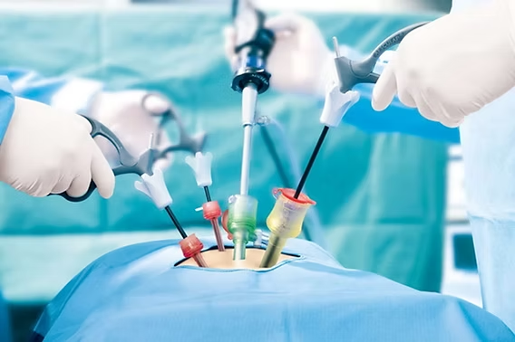
Understanding Gastrointestinal Surgical Conditions: Gastrointestinal surgeries encompass procedures involving the entire digestive tract, from esophagus to rectum. These surgeries address both benign and malignant conditions affecting your digestive health and overall quality of life.
Common Conditions Requiring GI Surgery:
- Inflammatory bowel disease (IBD)
- Colorectal cancer and polyps
- Gastric and duodenal ulcers
- Bowel obstructions
- Diverticulitis complications
- Appendicitis and peritonitis
When to Seek Our Expert GI Surgical Care:
Immediate Attention Required If You Experience:
- Severe abdominal pain with fever
- Persistent vomiting and inability to keep food down
- Blood in stool or vomit
- Sudden onset severe abdominal pain
- Signs of bowel obstruction
| Symptom Category | Description | Action Required |
|---|
| Early Signs | Persistent abdominal discomfort, changes in bowel habits | Schedule consultation |
| Progressive Symptoms | Chronic pain, unexplained weight loss, fatigue | Urgent evaluation needed |
| Acute Complications | Severe pain + fever + vomiting | Emergency visit required |
| Critical Signs | Peritonitis signs + shock + severe distension | IMMEDIATE EMERGENCY |
Our Advanced GI Surgery Approach:
🔬 Precise Diagnosis:
- CT and MRI imaging for detailed visualization
- Colonoscopy and upper endoscopy procedures
- Advanced tumor markers and genetic testing
- Multidisciplinary team evaluation
⚕️ Minimally Invasive Surgical Options:
- Laparoscopic colon and rectal surgeries
- Single-incision laparoscopic surgery (SILS)
- Robotic-assisted colorectal procedures
- Endoscopic mucosal resection (EMR)
🏥 Comprehensive Post-Surgical Care:
- Enhanced recovery after surgery (ERAS) protocols
- Specialized nutrition support
- Pain management with multimodal approach
Why choose Primax Hospital?
Our surgical team specializes in advanced minimally invasive techniques and provides personalized care throughout your treatment journey.
With state-of-the-art technology, expert surgeons, and a dedicated support staff, we ensure safe, effective, and compassionate care for every patient undergoing laparoscopic small bowel surgery.
Hernia Surgeries – Expert Repair with Advanced Techniques
Hernias affect millions of people worldwide, but with our specialized hernia surgery expertise, you can achieve permanent relief with minimal downtime and excellent cosmetic results.
Understanding Hernia Conditions: A hernia occurs when an internal organ pushes through a weak spot in your muscle or tissue wall. While some hernias cause minimal discomfort, others can lead to serious complications requiring immediate surgical intervention.
Types of Hernias We Treat:
- Inguinal hernias (groin area)
- Umbilical hernias (belly button)
- Incisional hernias (previous surgical sites)
- Hiatal hernias (diaphragm area)
- Ventral and epigastric hernias
Recognition and Symptoms Guide:
| Hernia Type | Key Symptoms | Surgical Urgency |
|---|
| Inguinal | Groin bulge, lifting discomfort | Elective planning |
| Umbilical | Naval protrusion, tender area | Schedule consultation |
| Incisional | Bulge at surgical site, pain | Early intervention |
| Strangulated | Severe pain + nausea + fever | EMERGENCY SURGERY |
When to Seek Our Hernia Surgery Expertise:
Immediate Emergency Signs:
- Sudden severe pain at hernia site
- Hernia becomes hard and cannot be pushed back
- Nausea and vomiting with hernia pain
- Fever with hernia complications
- Changes in skin color over the hernia
Our Advanced Hernia Surgery Techniques:
🔧 Minimally Invasive Approaches:
- TEP (Totally Extraperitoneal) repair
- Laparoscopic inguinal hernia repair
- Robotic-assisted complex hernia repairs
- Single-incision laparoscopic techniques
🧵 Advanced Mesh Technologies:
- Bio-compatible synthetic meshes
- Composite meshes for complex cases
- Absorbable tack fixation systems
- Tension-free repair techniques
⚡ Benefits of Our Hernia Surgery:
- Same-day discharge for most procedures
- Minimal post-operative pain
- Faster return to physical activities
- Lower recurrence rates (< 1%)
- Excellent cosmetic outcomes
Hepatobiliary and Pancreatic Surgeries – Complex Organ System Excellence
The hepatobiliary and pancreatic system requires the most sophisticated surgical expertise. Our specialized team handles complex liver, gallbladder, bile duct, and pancreatic surgeries with exceptional precision and outcomes.
Understanding HPB Surgical Conditions: Hepatobiliary and pancreatic surgeries involve some of the most complex procedures in general surgery, requiring extensive expertise and state-of-the-art facilities for optimal patient outcomes.
Conditions We Treat:
- Gallbladder stones and cholecystitis
- Liver tumors and cysts
- Pancreatic cancer and cysts
- Bile duct disorders
- Portal hypertension
- Chronic pancreatitis
Diagnostic and Symptom Recognition:
| Condition Category | Primary Symptoms | Diagnostic Priority |
|---|
| Gallbladder Disease | Right upper quadrant pain after meals | Routine evaluation |
| Liver Pathology | Jaundice + abdominal swelling + fatigue | Urgent assessment |
| Pancreatic Issues | Severe back/abdominal pain + weight loss | Immediate evaluation |
| Bile Duct Problems | Progressive jaundice + dark urine + pale stools | EMERGENCY WORKUP |
When to Consult Our HPB Surgery Specialists:
Immediate attention indicators:
- Progressive jaundice with fever
- Severe upper abdominal pain radiating to back
- Unexplained weight loss with abdominal pain
- New-onset diabetes with abdominal symptoms
- Abnormal liver function tests
Our Advanced HPB Surgery Capabilities:
🏥 Complex Surgical Procedures:
- Whipple procedure (pancreaticoduodenectomy)
- Liver resections and transplant evaluation
- Laparoscopic cholecystectomy with SILS technique
- Hepaticojejunostomy for bile duct reconstruction
- Pancreatic enucleation procedures
🔬 Advanced Diagnostic Integration:
- ERCP (Endoscopic Retrograde Cholangiopancreatography)
- EUS (Endoscopic Ultrasound) for precise staging
- Advanced MRI with MRCP protocols
- Intraoperative ultrasound guidance
✨ Specialized Care Features:
- Multidisciplinary tumor board discussions
- Enhanced recovery protocols for major surgeries
- Nutritional support and diabetes management
- Long-term follow-up with oncology integration
Why Choose Our Hospital?
Our hospital is renowned for its expertise in laparoscopic liver surgery, providing high-quality care with advanced diagnostic tools and modern surgical techniques.
Our skilled surgical team ensures precise, safe removal of liver cysts with a focus on minimal disruption to surrounding tissues.
We provide the best possible treatment outcomes with personalized care, shorter recovery times, and a commitment to patient well-being. Choose us for expert care and a smooth recovery experience.
Breast and Endocrine Surgeries – Specialized Hormonal and Breast Health Care
Our breast and endocrine surgery division provides comprehensive surgical care for breast conditions and endocrine disorders, combining surgical excellence with compassionate patient-centered care.
Understanding Breast and Endocrine Surgical Needs: Breast and endocrine surgeries require specialized expertise in handling delicate hormonal balance and breast tissue, with emphasis on both functional outcomes and cosmetic considerations.
Conditions We Expertly Treat:
- Breast cancer (all stages)
- Benign breast lumps and cysts
- Thyroid nodules and cancer
- Parathyroid disorders
- Adrenal gland tumors
- Gynecomastia (male breast enlargement)
Symptom Recognition and Action Guide:
| Condition Type | Warning Signs | Recommended Action |
|---|
| Breast Changes | New lump, skin changes, nipple discharge | Within 2 weeks consultation |
| Thyroid Issues | Neck swelling, voice changes, difficulty swallowing | Prompt evaluation |
| Hormonal Symptoms | Unexplained weight changes, energy fluctuations | Comprehensive assessment |
| Emergency Signs | Rapid growth, severe pain, systemic symptoms | IMMEDIATE EVALUATION |
When to Seek Our Breast & Endocrine Surgery Expertise:
Prompt Consultation Needed For:
- Any new breast lump or thickening
- Changes in breast size or shape
- Nipple discharge or skin changes
- Thyroid nodules or neck masses
- Family history of breast/ovarian cancer
- Abnormal hormone levels requiring surgical intervention
Our Advanced Surgical Approaches:
🎯 Minimally Invasive Techniques:
- Sentinel lymph node biopsy
- Laparoscopic thyroidectomy
- Endoscopic parathyroidectomy
- Vacuum-assisted breast biopsy
🔬 Precision Surgical Options:
- Breast-conserving surgery with oncoplastic reconstruction
- Total thyroidectomy with nerve monitoring
- Robotic-assisted endocrine surgeries
- Immediate breast reconstruction coordination
💝 Comprehensive Care Features:
- Genetic counseling and testing
- Multidisciplinary cancer care team
- Psychosocial support services
- Cosmetic outcome optimization
- Long-term survivorship planning


 Comprehensive GI Surgical Expertise

