
Primax Neurosurgery Center: Advanced Brain, Spine & Neuro Trauma Care in Sonipat
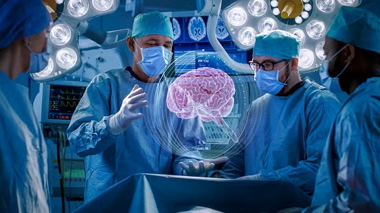
Welcome to the Neurosurgery Department at Primax Gastro Institute & Superspeciality Hospital, Sonipat, where advanced neurological care meets compassionate healing. Our world-renowned neurosurgery team delivers exceptional outcomes for complex brain surgery, spine surgery, and neurotrauma cases using state-of-the-art technology and minimally invasive techniques.
As the best neurosurgeon in Sonipat, our specialists combine decades of surgical expertise with cutting-edge neuro-navigation systems to treat conditions ranging from brain tumors to head injury cases with precision and care.
At Primax Neurosurgery Center, we understand that neurological conditions require not just surgical excellence but also empathetic patient care. Our multidisciplinary approach ensures comprehensive treatment from diagnosis through recovery, making us the premier destination for neurosurgery in Haryana.
Whether you’re dealing with spinal disc herniation, brain hemorrhage, or any complex neurological disorder, our 24/7 emergency neurosurgery services stand ready to provide life-saving care when every second counts.
✅ World-Class Neurosurgical Expertise
✅ State-of-the-Art Neuro-Surgical Infrastructure
✅ Minimally Invasive Neurosurgery
✅ Comprehensive Neuro-Care Services
Brain Tumors (Benign and Malignant) – Expert Brain Surgery in Sonipat
Brain tumors can be life-altering, but with expert neurosurgery care, many patients achieve excellent outcomes. Our brain tumor specialists in Sonipat utilize advanced surgical techniques and neuro-oncology protocols to provide comprehensive treatment for all types of brain tumors.
Brain tumors are abnormal growths of cells within the brain or skull. They can be benign (non-cancerous) or malignant (cancerous), and both types require expert evaluation and treatment. At Primax Neurosurgery Center, we provide compassionate, expert care throughout your journey.
Common Types of Brain Tumors We Treat:
Recognizing Brain Tumor Symptoms
| Symptom Category | Description | Action Required |
|---|---|---|
| Early Warning Signs | Persistent headaches, vision changes, mild confusion | Schedule consultation |
| Progressive Symptoms | Seizures, personality changes, weakness in limbs | Urgent evaluation needed |
| Severe Symptoms | Severe headache, vomiting, loss of consciousness | Emergency visit |
| Critical Signs | Sudden seizure, stroke-like symptoms, severe confusion | IMMEDIATE EMERGENCY |
When to Seek Our Expert Neurosurgery Care
Our Advanced Treatment Approach for Brain Tumors
A. Precise Diagnosis:
B. Comprehensive Treatment Options:
C. Integrated Cancer Care:
Why Choose Primax Neurosurgery Center for Brain Tumor Treatment?
🏃 Take the Next Step Toward Recovery
If you or a loved one has been diagnosed with a brain tumor, early expert intervention improves outcomes.
Consult our neurosurgery specialists at Primax Gastro Institute & Super Speciality Hospital – Call 9666460009 or visit our Contact Us page to schedule a comprehensive evaluation.
Spinal Disc Herniation (Slip Disc) – Advanced Spine Surgery Solutions
Slipped disc or spinal disc herniation is one of the most common causes of back pain and leg pain. Our spine surgery specialists in Sonipat offer both conservative and advanced surgical solutions to help you return to an active, pain-free life.
A herniated disc occurs when the soft inner core of a spinal disc pushes through a tear in the outer layer, often compressing nearby nerves. This condition, commonly called a “slip disc,” can cause severe pain, numbness, and weakness. At Primax Hospital, we provide comprehensive evaluation and treatment options tailored to your specific condition.
Common Causes of Disc Herniation:
Recognizing Slip Disc Symptoms
| Symptom Category | Description | Action Required |
|---|---|---|
| Mild Symptoms | Occasional back pain, slight stiffness | Conservative management, schedule check-up |
| Moderate Symptoms | Radiating leg pain (sciatica), numbness in feet | Consultation within 1 week |
| Severe Symptoms | Severe pain limiting movement, weakness in legs | Urgent evaluation needed |
| Emergency Signs | Loss of bladder/bowel control, saddle anesthesia | IMMEDIATE EMERGENCY SURGERY |
When to Seek Our Spine Surgery Expertise
Our Advanced Treatment Approach for Slipped Disc
A. Comprehensive Diagnosis:
B. Conservative Treatment Options:
C. Advanced Surgical Interventions:
Why Choose Primax Hospital for Spine Surgery?
🏃 Get Relief from Chronic Back Pain
Don’t let slip disc control your life. Contact our spine surgery specialists at Primax Gastro Institute & Super Speciality Hospital – Call 9666460009 or book your appointment online for a comprehensive spine evaluation.
Spinal Cord Compression – Emergency Neurosurgery Care
Spinal cord compression is a serious neurological emergency that requires immediate expert intervention. Our neurosurgery team provides 24/7 emergency care for spinal cord compression cases, ensuring the best possible outcomes through rapid diagnosis and treatment.
Spinal cord compression occurs when abnormal pressure is placed on the spinal cord, the highway of nerves connecting your brain to the rest of your body. This pressure can result from various causes and can lead to permanent neurological damage if not treated promptly. At Primax Hospital, we treat spinal cord compression as the medical emergency it is.
Common Causes We Treat:
Critical Symptom Recognition Guide
| Symptom Category | Description | Action Required |
|---|---|---|
| Early Warning | Mild weakness, altered sensation in limbs | Urgent consultation within 24 hours |
| Progressive Signs | Increasing weakness, difficulty walking, bowel/bladder changes | SAME-DAY EMERGENCY VISIT |
| Severe Symptoms | Unable to walk, severe weakness in all limbs, loss of sensation | IMMEDIATE EMERGENCY |
| Complete Compression | Paralysis, complete loss of sensation below level, incontinence | CRITICAL – CALL AMBULANCE |
When to Seek Emergency Neurosurgery Care
Our Emergency Treatment Protocol
A. Rapid Diagnosis (Within 30 Minutes):
B. Immediate Medical Stabilization:
C. Emergency Surgical Interventions:
D. Post-Operative Rehabilitation:
Why Choose Primax for Spinal Cord Compression Emergencies?
❤️ Emergency Neurosurgery Contact
If you or someone you know is experiencing symptoms of spinal cord compression, call immediately at 9666460009 for emergency neurosurgery consultation. Don’t wait – permanent nerve damage can occur within hours.
Head Injuries and Trauma – 24/7 Neuro-Trauma Care in Sonipat
Head injuries require immediate expert evaluation and treatment. As the leading center for head injury treatment in Sonipat, Primax Neurosurgery Center provides comprehensive neuro-trauma care 24/7, from minor concussions to life-threatening traumatic brain injuries.
Head injuries range from mild concussions to severe traumatic brain injuries (TBI) that can be life-threatening. At Primax Hospital, we treat all severities of head injury with the urgency and expertise they deserve. Our neuro-trauma team is available 24/7 to provide immediate life-saving care.
Common Types of Head Injuries We Treat:
Head Injury Severity Assessment
| Severity Level | Symptoms | Management |
|---|---|---|
| Mild (Concussion) | Brief loss of consciousness, confusion, headache | Observation, rest, follow-up in 24-48 hours |
| Moderate | Prolonged confusion, vomiting, severe headache, drowsiness | CT scan, hospital admission, close monitoring |
| Severe | Unconsciousness >5 minutes, seizures, unequal pupils | EMERGENCY SURGERY, ICU care, ventilator support |
| Critical | Coma, no response, abnormal posturing | IMMEDIATE LIFE-SAVING SURGERY, Neuro-ICU |
When to Seek Emergency Care After Head Injury
Our Comprehensive Head Injury Treatment Protocol
A. Emergency Assessment (Golden Hour):
B. Medical Management:
C. Emergency Neurosurgical Interventions:
D. Neuro-Rehabilitation:
Why Choose Primax for Head Injury Treatment in Sonipat?
❤️ Emergency Head Injury Contact
For head injury emergencies, call our 24/7 emergency line at 9666460009 immediately. In severe cases, ask for an ambulance. Time matters in brain injury – we’re ready to save lives around the clock.
Hydrocephalus – Expert Diagnosis and Shunt Surgery
Hydrocephalus, often called “water on the brain,” requires specialized neurosurgical expertise. Our team provides comprehensive care for all types of hydrocephalus, from newborns to adults, using the latest surgical techniques and shunt technology.
Hydrocephalus is a condition where excess cerebrospinal fluid (CSF) accumulates in the brain’s ventricles, causing increased pressure that can damage brain tissue. At Primax Neurosurgery Center, we specialize in treating all forms of hydrocephalus with both traditional shunt surgery and advanced endoscopic techniques.
Types of Hydrocephalus We Treat:
Recognizing Hydrocephalus Symptoms by Age
| Age Group | Key Symptoms | Action Required |
|---|---|---|
| Infants | Rapid head growth, bulging fontanel, vomiting, sleepiness | Urgent pediatric neurosurgery consultation |
| Children | Headache, vision problems, developmental delays, poor school performance | Schedule evaluation within 1 week |
| Adults | Walking difficulty, urinary incontinence, memory problems | Consultation needed |
| Emergency Signs | Severe headache, vomiting, altered consciousness, seizures | IMMEDIATE EMERGENCY VISIT |
When to Seek Specialized Care
In Infants:
In Older Children and Adults:
Our Advanced Treatment Approach
A. Comprehensive Diagnosis:
B. Surgical Treatment Options:
C. Medical Management:
Why Choose Primax for Hydrocephalus Treatment?
❤️ Schedule Your Hydrocephalus Consultation
Early treatment prevents brain damage and improves long-term outcomes. Contact our neurosurgery specialists at Primax Gastro Institute & Super Speciality Hospital at 9666460009 or book online for expert evaluation.
Brain Hemorrhage / Intracranial Bleeds – Life-Saving Emergency Neurosurgery
Brain hemorrhage is a life-threatening emergency that demands immediate expert neurosurgical intervention. Our 24/7 stroke and hemorrhage response team provides rapid diagnosis and treatment, significantly improving survival and recovery outcomes.
Brain hemorrhage, also called intracranial bleeding, occurs when a blood vessel in or around the brain ruptures, causing blood to leak into brain tissue or surrounding spaces. This creates pressure that can damage brain cells and threaten life. At Primax Neurosurgery Center, we treat brain hemorrhage as the critical emergency it is, with our team ready to intervene at any hour.
Types of Intracranial Bleeding We Treat:
Emergency Symptom Recognition
| Symptom Severity | Presentation | Required Action |
|---|---|---|
| Moderate Bleed | Severe headache, vomiting, and confusion | Emergency department visit NOW |
| Large Bleed | Severe headache, weakness, speech difficulty, vision loss | CALL AMBULANCE IMMEDIATELY |
| Massive Bleed | Loss of consciousness, seizures, abnormal breathing | CRITICAL – 911/EMERGENCY SERVICES |
Warning Signs: “Worst headache of life”, sudden severe headache with vomiting – do not delay, EMERGENCY VISIT required.
When to Call Emergency Services
Our Emergency Brain Hemorrhage Protocol
A. Ultra-Rapid Response (Within 15 Minutes):
B. Immediate Medical Stabilization:
C. Emergency Neurosurgical Procedures:
D. Intensive Neuro-Critical Care:
Why Choose Primax for Brain Hemorrhage Emergencies?
❤️ Brain Hemorrhage Emergency Contact
Brain hemorrhage is a race against time. Call 9666460009 immediately if you suspect a brain bleed or book online. Our neuro-emergency team is standing by 24/7. Don’t wait – every minute counts in saving brain tissue and lives.
Trigeminal Neuralgia – Advanced Pain Management and Surgical Solutions
Trigeminal neuralgia, often described as the “suicide disease” due to its excruciating pain, doesn’t have to control your life. Our neurosurgery team specializes in both medical and surgical treatments that can provide lasting relief from this debilitating facial pain condition.
Trigeminal neuralgia is a chronic pain condition affecting the trigeminal nerve, which carries sensation from your face to your brain. Even mild stimulation of your face can trigger jolts of excruciating pain. At Primax Hospital, we understand how this condition can devastate quality of life, and we’re here to help you find relief.
Common Triggers and Causes:
Pain Pattern Recognition
| Pain Characteristic | Description | Impact Level |
|---|---|---|
| Typical TN | Electric shock-like, seconds to minutes, triggered by touch/chewing | Severe – Limits eating, talking, face washing |
| Atypical TN | Constant burning, aching pain with occasional sharp attacks | Moderate to Severe – Affects quality of life |
| TN2 | Continuous background pain with breakthrough sharp pain | Severe – Often treatment resistant |
| Symptomatic TN | Associated with other neurological symptoms | Variable – May indicate underlying condition |
When to Seek Specialized Neurosurgery Care
Our Comprehensive Treatment Approach
A. Accurate Diagnosis:
B. Medical Management:
C. Advanced Surgical Solutions:
Why Choose Primax for Trigeminal Neuralgia Treatment?
🏃 Get Relief from Facial Pain
Don’t suffer in silence. Trigeminal neuralgia is treatable. Contact our neurosurgery specialists at Primax Gastro Institute & Super Speciality Hospital at 9666460009 or schedule your consultation online to explore your treatment options.
Cervical and Lumbar Spondylosis – Advanced Spine Degeneration Treatment
Cervical and lumbar spondylosis are common age-related degenerative conditions of the spine that can cause chronic pain and disability. Our spine surgery specialists offer comprehensive treatment from conservative management to advanced surgical solutions, helping you maintain an active, pain-free lifestyle.
Spondylosis is wear-and-tear arthritis affecting the spine’s bones, discs, and joints. Cervical spondylosis affects the neck, while lumbar spondylosis affects the lower back. At Primax Neurosurgery Center, we understand that while spondylosis is common with aging, the pain and limitations it causes are not something you have to accept.
Common Causes and Risk Factors:
Symptom Recognition by Location
| Condition | Primary Symptoms | Advanced Symptoms | Action Required |
|---|---|---|---|
| Cervical Spondylosis | Neck pain, stiffness, headaches | Arm pain, numbness, weakness in hands | Consultation if symptoms persist >2 weeks |
| Lumbar Spondylosis | Low back pain, morning stiffness | Leg pain (sciatica), difficulty walking | Evaluation if affecting daily activities |
| Cervical Myelopathy | Difficulty with fine hand movements, unsteady gait | Severe weakness, bowel/bladder issues | URGENT – Can lead to paralysis |
| Cauda Equina Syndrome | Severe low back pain, leg weakness | Loss of bowel/bladder control, saddle numbness | EMERGENCY SURGERY REQUIRED |
When to Seek Spine Surgery Consultation
Our Comprehensive Treatment Protocol
A. Advanced Diagnostic Evaluation:
B. Conservative Management (First-Line Treatment):
C. Surgical Solutions (When Conservative Treatment Fails):
Why Choose Primax for Spondylosis Treatment?
🏃 Start Your Journey to Pain Relief
Don’t let spine degeneration limit your life. Contact our spine specialists at Primax Gastro Institute & Super Speciality Hospital at 9666460009 or book your consultation online for a comprehensive spine evaluation and personalized treatment plan.


Thank You for Submitting
You appointment query has been registered you will soon recieve confirmation call from Hospital if not received call@9666460009
