






If your doctor orders a scan, you want to know what it will show and why that test was picked. Both CT and MRI take pictures of your insides, but they use different tools and answer different questions.
Choose a CT scan when you need fast answers in an emergency, for bone fractures, bleeding, lung problems, or kidney stones, and choose an MRI when your doctor needs fine detail of soft tissue like the brain, spinal cord, joints, or a suspected tumor. That single difference explains most of the choices doctors make between the two.
Computed tomography uses X-rays and finishes in minutes. Magnetic resonance imaging uses a strong magnet and radio waves, takes longer, and shows soft tissue contrast that a CT cannot match. Neither test is “better” in general; they are tuned for different jobs.
By the end of this, you will be able to tell what your scan is looking for, what prep it needs, and what questions to ask before you walk into the room.
How Do CT and MRI Create Images?
A CT scanner spins an X-ray tube around your body and measures how much of the beam each tissue absorbs, then a computer builds cross-section slices from those measurements. StatPearls explains that CT images come from a series of X-rays generated by a rapidly rotating tube, and that CT costs less than MRI.
Because it uses X-rays, a CT scan involves ionizing radiation. The dose is small for a single study, and the machine only needs seconds of actual scanning time, which is why it works so well when someone is in pain or cannot hold still.
MRI takes a different path. A powerful magnet lines up water molecules in your body, short radio wave pulses knock them out of line, and the signal they give off as they settle back is turned into images.
No X-rays are involved. The Mayo Clinic notes that a brain CT uses X-rays and a computer to picture the brain and skull, while MRI relies on magnets and radio waves.
What does that mean for the pictures?
| Feature | CT | MRI |
| Energy used | X-rays | Magnets + radio waves |
| Radiation | Yes | None |
| Scan time | Minutes | 30 to 60 minutes |
| Best at | Bone, bleeding, lung | Soft tissue, brain, nerves |
| Noise level | Quiet | Loud knocking |
CT shows bones and fresh blood sharply. MRI separates one soft tissue from another, which is how it picks up small changes inside organs, muscle, and nerve tissue. Our radiology and imaging services team matches the machine to the question being asked.
When Is CT Usually the Better Choice?
CT is the first test in nearly every emergency, because it is fast, and speed changes outcomes. In a trauma room, a full body CT can be done while the patient is still on the stretcher.
Doctors reach for CT in these situations:
- Injuries and fractures. Bone fractures, skull injuries, and spine breaks show up clearly.
- Internal bleeding. Fresh blood looks bright on CT, so it is the standard first scan for suspected bleeding in the head or belly.
- Stroke. A quick head CT tells the team whether a stroke is caused by a clot or a bleed, which decides the treatment.
- Lung problems. A chest CT finds pneumonia, fluid, and scarring. Low-dose chest CT is also used for lung cancer screening in heavy smokers.
- Kidney stones. CT finds stones of nearly any size and shows exactly where they sit.
- Abdominal emergencies. Appendicitis, bowel obstruction, and abscesses are all CT territory.
- Blood vessels. CT angiography (CTA) maps blood vessels to look for pulmonary embolism, blood clots, and an abdominal aortic aneurysm.
- Cancer staging. CT of the chest, abdomen, and pelvis is the workhorse for checking whether cancer has spread.
CT also wins when a patient cannot lie still, has a pacemaker, or is on a ventilator. A study of musculoskeletal imaging found that CT better defines bone detail and calcification that MRI sequences read as nonspecific.
If you arrive at our emergency department with chest pain, belly pain, or a head injury, expect a CT before anything else.
When Does MRI Provide More Detail?
MRI earns its extra time whenever the question is about soft tissue. It can separate gray matter from white matter, healthy muscle from torn muscle, and a tumor from the normal tissue around it.
Brain and nerves. MRI finds small brain tumors, early multiple sclerosis plaques, and spinal cord compression that a CT can miss. Research on soft-tissue tumors reported that CT did not provide precise anatomic detail of lesions in the way MRI did. Complex brain and spine cases are often planned with our neurosurgery team using MRI images.
Joints. Cartilage, tendons, ligaments, and a torn meniscus are visible on MRI without any radiation. Knee, shoulder, and hip injuries handled by our orthopedics department almost always need MRI before surgery.
Tumors and masses. MRI shows how far a mass reaches into nearby tissue, which matters for planning surgery or radiation.
Blood vessels and heart. MR angiography maps vessels without X-rays. Cardiac MRI measures heart muscle scarring and pumping strength.
Abdomen. MRI is stronger than CT for characterizing liver lesions, and MRCP produces a detailed map of the bile ducts and pancreas ducts without a scope. Digestive problems seen in our gastroenterology clinic often move from ultrasound to MRI when the diagnosis is unclear. Kidney masses are also studied with MRI when contrast CT is risky.
The trade-off is time and comfort. You lie still for 30 to 60 minutes inside a narrow tube while the machine knocks loudly.
Safety, Contrast, and Preparation Considerations
Both scans have strong safety records, and the risks are different for each.Radiologists describe MRI and CT as indispensable imaging modalities with excellent safety profiles that still carry specific physical and procedural risks.
Radiation. A single CT gives a small dose. The concern is cumulative radiation from many scans over years, which raises a small theoretical cancer risk. That is why doctors avoid repeat CTs when MRI or ultrasound answers the question.
Pregnancy. MRI without contrast is preferred during pregnancy. When CT is needed for a serious emergency, the benefit can outweigh the risk, and the team will shield and limit the scan. Questions about imaging while expecting are worth raising with your doctor during pregnancy care visits.
Contrast dye. CT uses iodine-based contrast. MRI uses gadolinium-based contrast. These agents work by different mechanisms, iodine through X-ray attenuation and gadolinium through magnetic properties. Tell staff about any past allergic reaction.
Kidneys. Both dyes are filtered by the kidneys, so a blood test for kidney function (GFR) is often done first. Your results come through our laboratory services.
Metal and devices. Pacemakers, defibrillators, cochlear implants, insulin pumps, and some older metal implants can be unsafe in an MRI magnet. Bring device cards with you. Many modern pacemakers are MRI-conditional, but the model must be confirmed.
Claustrophobia. Tell the technologist before the scan. An open MRI, a mild sedative, or headphones and a blindfold all help.
What to Expect During the Scan and Afterward
You will change into a gown, remove metal, and lie on a table that slides into the machine.
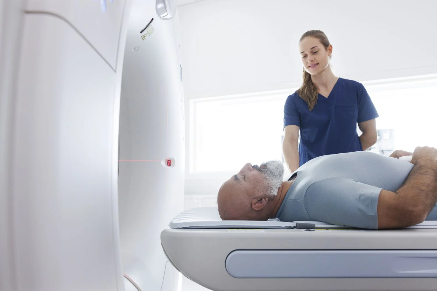
For a CT, the table moves through a wide, donut-shaped ring. The scan takes minutes, and you will hear soft whirring. You may be asked to hold your breath for a few seconds.
For an MRI, you lie in a longer tube. The machine knocks and buzzes loudly, so you get earplugs or headphones. Staying still is the single most important thing you do; motion blurs the images and can force a repeat.
If contrast dye is used, an IV goes in your arm. CT contrast often brings a warm flush and a metallic taste for a few seconds. Gadolinium is quieter, sometimes with a cool feeling at the IV site.
Afterward, you can eat and drive as normal unless you were sedated. Drink extra water that day to help clear the dye.
A radiologist reads the images and sends a report to your doctor, often the same day for urgent cases. Ask when and how you will get results before you leave the department.
If symptoms continue or get worse after a normal scan, come back. A follow-up visit with one of our doctors can decide whether a different test is needed.
Frequently Asked Questions
Is the radiation exposure from a CT scan dangerous?
A single CT scan carries a very small risk, and doctors order it when the information it gives outweighs that risk. The concern grows with many scans over many years, so tell your doctor about past CTs. MRI and ultrasound use no radiation and are chosen when they can answer the same question.
Can I have an MRI with a pacemaker or metal implants?
Many newer pacemakers and defibrillators are labeled MRI-conditional and can be scanned under specific settings, but the exact model must be checked first. Older devices, cochlear implants, and some metal fragments can rule out MRI. Bring your device card and implant paperwork to your appointment.
Which scan is better for kidney stones?
CT is the preferred test for kidney stones because it shows stone size, number, and exact position within minutes. Ultrasound is used first for children and pregnant patients. MRI does not show most stones well.
Do I need a CT or MRI for back and spine pain?
MRI is the better test for most back pain that involves nerve symptoms, because it shows discs, the spinal cord, and nerve roots. CT is chosen after trauma, when a fracture is the main concern, or when MRI cannot be done. Many cases of simple back pain need no scan at all.
Why does MRI show soft tissues so much better than CT?
MRI reads signals from water and fat in tissue, so small differences between two soft tissues create visible contrast. CT measures X-ray absorption, which is similar across most soft tissues but very different for bone and blood. That is why the two tests are used for different questions.
Where can I get a CT or MRI done in Sonipat?
Primax Hospital on Bahalgarh Road, Sector-4, Sonipat-131001, Haryana runs CT and MRI services with in-house radiologists, and patients from Sonipat and across Haryana are seen daily. Walk in or book ahead if your symptoms are not improving, especially after an injury, ongoing headaches, belly pain, or joint pain.
Call +91 96664 60009 to schedule your scan or speak with our imaging team.

